Every year I watch new NPs order an echocardiogram to answer a question the physical exam would have answered in 30 seconds.
Not because they’re lazy. Because nobody taught them to use their hands.
What Got Lost
The physical exam was never just a ritual. It was a diagnostic tool — and in cardiology, it is still one of the most information-dense tools available. The jugular venous pressure tells you about right heart filling pressures. The quality of the carotid upstroke tells you about aortic valve function. The position and character of the PMI tells you about ventricular remodeling. The presence or absence of an S3 tells you more about volume status than a BNP in many clinical contexts.
None of this requires technology. It requires training.
What happened is that training programs across medicine deprioritized bedside examination skills as imaging became faster and more accessible. Why spend an hour teaching students to assess JVP when you can just get a CVP? Why teach palpation of the liver for hepatomegaly when ultrasound is in the room?
The answer is that imaging answers the question you thought to ask. The physical exam surfaces findings you didn’t know to look for.
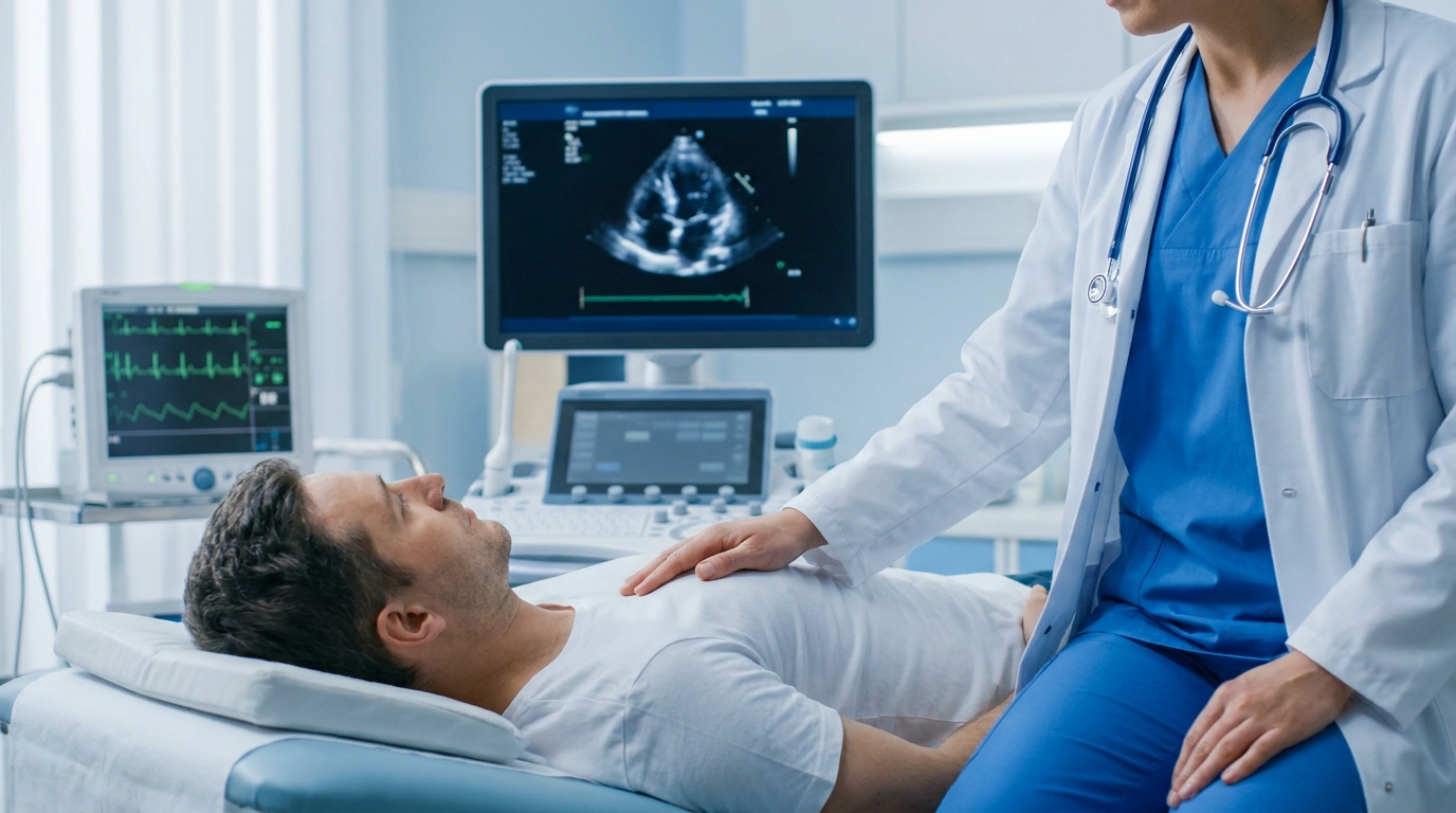
The Cardiology Case for Bedside Examination
In acute care cardiology, the physical exam is not supplementary. It is often the fastest way to triage a deteriorating patient.
A patient with worsening dyspnea: Is the JVP elevated? Are there crackles at the bases? Is there a new S3? If yes to all three, you’re looking at decompensated heart failure and you can start treatment before the BNP comes back. If the JVP is flat and the lungs are clear, heart failure is less likely and you need to think differently.
That decision takes 90 seconds with trained hands. It takes two hours waiting on labs and imaging.
A patient with a new murmur post-MI: Is it holosystolic, harsh, and loudest at the apex? Does it radiate to the axilla? That’s mitral regurgitation, probably papillary muscle dysfunction. Is it holosystolic at the lower sternal border with a thrill? That’s a VSD until proven otherwise and it’s a different emergency entirely. The physical exam told you which before the echo was even ordered.
What Good Teaching Looks Like
The students who develop strong exam skills have almost always had one thing in common: someone made them do it slowly, repeatedly, and without the safety net of imaging available.
Not once in a simulation lab. Not a checklist. Actually examining patients, with feedback, in clinical environments where the diagnosis was later confirmed or refuted by imaging. The correlation between what you felt and what the echo showed is how you build the neural map that makes physical examination useful.
Programs that gut clinical hours in favor of didactic content, or that accept documentation of clinical encounters without direct supervision, produce graduates who technically completed a physical exam curriculum and cannot perform a reliable cardiac exam.
This is fixable. It requires clinical faculty who prioritize it and preceptors who model it in practice. It requires programs to stop treating the physical exam as a competency to document and start treating it as a skill to develop.
The technology will keep improving. The exam will still tell you things the technology misses. That gap doesn’t close — it grows, because technology optimizes for the diagnoses we already know to look for. The trained clinician looks for the ones nobody thought to order a test for.
Teach the exam. Teach it seriously. Patients benefit directly.